Title of Presentation
“Cruising inside cells”
The behavior of biochemical molecules moving around in cells makes me think of a school of whales wandering in the ocean, captured by the Argus system on the artificial satellite. When bringing a whale back into the sea — with a transmitter on its dorsal fin, every staff member hopes that it will return safely to a school of its species. A transmitter is now minute in size, but it was not this way before. There used to be some concern that a whale fitted with a transmitter could be given the cold shoulder and thus ostracized by other whales for “wearing something annoying.” How is whale’s wandering related to the tide or a shoal of small fish? What kind of interaction is there among different species of whales? We Human Beings have attempted to fully understand this fellow creature in the sea both during and since the age of whale fishing.
In a live cell imaging experiment, a luminescent probe replaces a transmitter. We label a luminescent probe on a specific region of a biological molecule and bring it back into a cell. We can then visualize how the biological molecule behaves in response to external stimulation. Since luminescence is a physical phenomenon, we can extract various kinds of information by making full use of its characteristics.
Cruising inside cells in a supermicro corps, gliding down in a microtubule like a roller coaster, pushing our ways through a jungle of chromatin while hoisting a flag of nuclear localization signal — we are reminded to retain a playful and adventurous perspective at all times. What matters is mobilizing all capabilities of science and giving full play to our imagination. We believe that serendipitous findings can arise out of such a sportive mind, a frame of mind that prevails when enjoying whale-watching.
Over the past two decades, various genetically encoded probes have been generated principally using fluorescent proteins. I will discuss how the probes have advanced our understanding of the spatio-temporal regulation of biological functions, such as cell-cycle progression, autophagy, and metabolism, inside cells, neurons, embryos, and brains. I will also speculate on how these approaches will continue to improve due to the various features of fluorescent proteins.
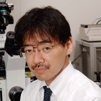
Profile
- A brief Biography
-
Mar. 1987 M.D., Keio University School of Medicine Mar. 1991 Ph.D., Osaka University Graduate School of Medicine (Molecular Neurobiology)
Research field: The molecular basis of the calcium channel activity of IP3 receptorsApr. 1991 Research Fellow, Japan Society for the Promotion of Science
Research field: Structure-function relationships of IP3 receptorsApr. 1993 – Dec. 1998 Assistant Professor, Institute of Medical Science, The University of Tokyo
Research field: IP3/calcium dynamicsOct. 1995 HFSP long-term fellowship, University of California San Diego, Dept. of Pharmacology
Research field: Development of a calcium probeOct. 1997 Research Pharmacologist, University of California San Diego, Dept. of Pharmacology
Research field: Development of a calcium probeJan. 1999 – Laboratory Head, Laboratory for Cell Function Dynamics, Advanced Technology Development Group, RIKEN Brain Science Institute
Research field: Fluorescent bioimagingJan. 2004 – Mar. 2009 Group Director, Advanced Technology Development Group, RIKEN Brain Science Institute
Research field: Fluorescent bioimagingJul. 2005 – Mar. 2010 Visiting Professor, Department of Proteomics, Research Center for Bioinformatics, Institute of Molecular and Cellular Biosciences, The University of Tokyo Apr. 2006 – Mar. 2011 Visiting Professor, Laboratory of Developmental Dynamics, National Institute for Basic Biology, The National Institute of Natural Sciences Oct. 2006 – Mar. 2012 Research Director, ERATO Life Function Dynamics Project, Japan Science and Technology Agency Apr.2007 – Visiting Professor, Department of Molecular Neurobiology, Graduate School of Advanced Science and Engineering, Waseda University Apr. 2008 – Deputy Director, RIKEN Brain Science Institute Apr. 2009 – Visiting Professor, Keio University School of Medicine Apr. 2010 – Mar. 2011 Visiting Professor, Faculty of Science, Toho University Apr. 2012 – Visiting Professor, Graduate School of Nanobioscience, Yokohama City University Apr. 2013 – Laboratory Head, Biotechnological Optics Research Team, RIKEN Center for Advanced Photonics - Details of selected Awards and Honors
-
Oct. 2004 Yamazaki-Teiichi Prize, Foundation for Promotion of Material Science and Technology of Japan (Biological Science & Technology) Dec. 2006 JSPS (Japan Society for the Promotion of Science) Prize (Biological Sciences) Mar. 2007 Tsukahara Memorial Award Jun. 2007 Kitasato Award, Keio University School of Medicine Alumni Association Apr. 2008 Prize for Science and Technology (Development Category), The Commendation for Science and Technology by the Minister of Education, Culture, Sports, Science and Technology, Feb. 2012 Inoue Prize for Science Jun. 2013 Fujihara Award Dec. 2014 Arthur Kornberg Memorial Award Oct. 2015 W. Alden Spencer Award (Columbia University) Feb. 2016 Shimadzu Award - A list of selected Publications
-
Hama H, Hioki H, Namiki K, Hoshida T, Kurokawa H, Ishidate F, Kaneko T, Akagi T, Saito T, Saido T, Miyawaki A. (2015) ScaleS: an optical clearing palette for biological imaging. Nature Neuroscience, 18: 1518-1529.
Kumagai A, Ando R, Miyatake H, Greimel P, Kobayashi T, Hirabayashi Y, Shimogori T, Miyawaki A. (2013) A Bilirubin-Inducible Fluorescent Protein from Eel Muscle. Cell, 153: 1602-1611.
Shimozono S, Iimura T, Kitaguchi T, Higashijima SI, Miyawaki A. (2013) Visualization of an endogenous retinoic acid gradient across embryonic development. Nature, 496: 363-366.
Hama H, Kurokawa H, Kawano H, Ando R, Shimogori T, Noda H, Fukami K, Sakaue-Sawano A, Miyawaki A. (2011) Scale: a chemical approach for fluorescence imaging and reconstruction of transparent mouse brain. Nature Neuroscience, 14: 1481-1488.
Sakaue-Sawano A, Kurokawa H, Morimura T, Hanyu A, Hama H, Osawa H, Kashiwagi S, Fukami K, Miyata T, Miyoshi H, Imamura T, Ogawa M, Masai H, Miyawaki A. (2008) Visualizing Spatiotemporal Dynamics of Multicellular Cell Cycle Progression. Cell, 132: 487-498.
Ando R, Mizuno H, Miyawaki A. (2004) Regulated fast nucleocytoplasmic shuttling observed by reversible protein highlighting. Science, 306: 1370-1373.
Mizuno H, Mal TK, Tong KI, Ando R, Furuta T, Ikura M, Miyawaki A. (2003) Photo-induced peptide cleavage in the green-to-red conversion of a fluorescent protein. Mol. Cell, 12: 1051-1058.
Ando R, Hama H, Yamamoto-Hino M, Mizuno H, Miyawaki A. (2002) An optical marker based on the UV-induced green-to-red photoconversion of a fluorescent protein. Proc. Natl. Acad. Sci. USA., 99: 12651-12656.
Nagai T, Ibata K, Park ES, Kubota M, Mikoshiba K, Miyawaki A. (2002) A variant of yellow fluorescent protein with fast and efficient maturation for cell-biological applications. Nature Biotechnology, 20: 87-90.
Miyawaki A, Llopis J, Heim R, McCaffery JM, Adams JA, Ikura M, Tsien RY. (1997) Fluorescent indicators for Ca2+ based on green fluorescent proteins and calmodulin. Nature, 388: 882-887.


